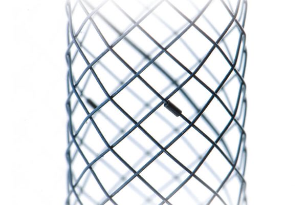
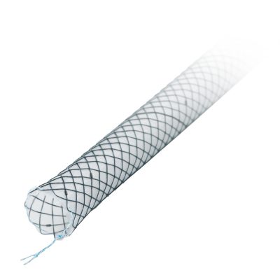
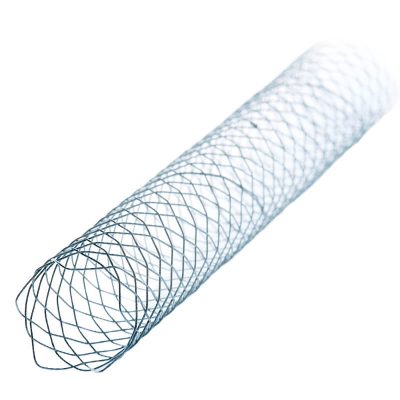
With the biliary stents manufactured by MICRO-TECH you will always make the ideal choice to bridge-graft biliary duct stenoses. The high expansion force of the Nitinol wire mesh accounts for an excellent positional stability. The resistant cover prevents the stent from growing into the tissue. The product range comprises two stent lines and offers a first-class solution that suits all requirements: the classic line and the platinum line. The comprehensive platinum line is especially distinguished by its extraordinary high radiopacity.
BILIARY DUCT STENTS
ALWAYS THE IDEAL SOLUTION
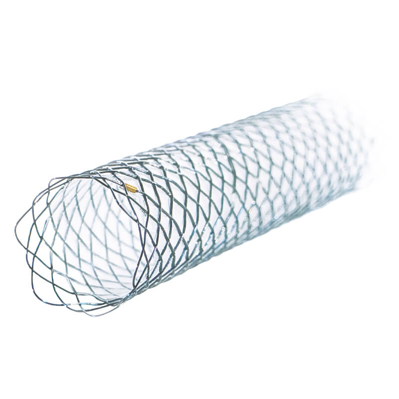
SPECIFIC CHARACTERISTICS
- Self-expanding
- Available for TTS and PTCD
- Classic- and Platinum-Line
- Nitinol wire mesh with atraumatic ends
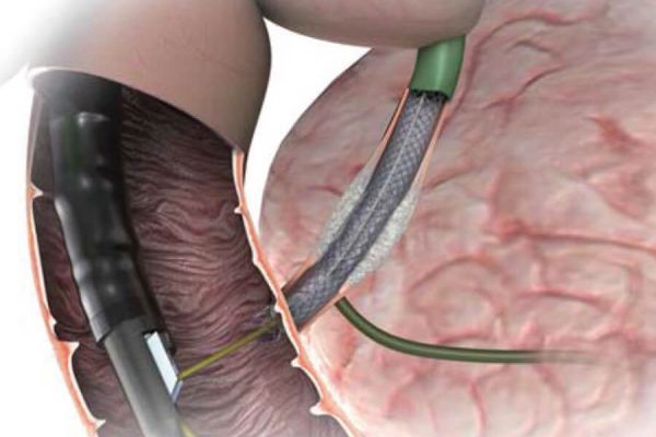
- Release under visual endoscopic control
- Enormous position stability
- Resistant and elastic cover
- High radiopacity
- Guiding wire passable to a maximum of 0.035 inches
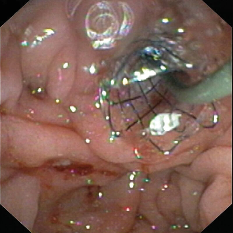
Removal of the introducer
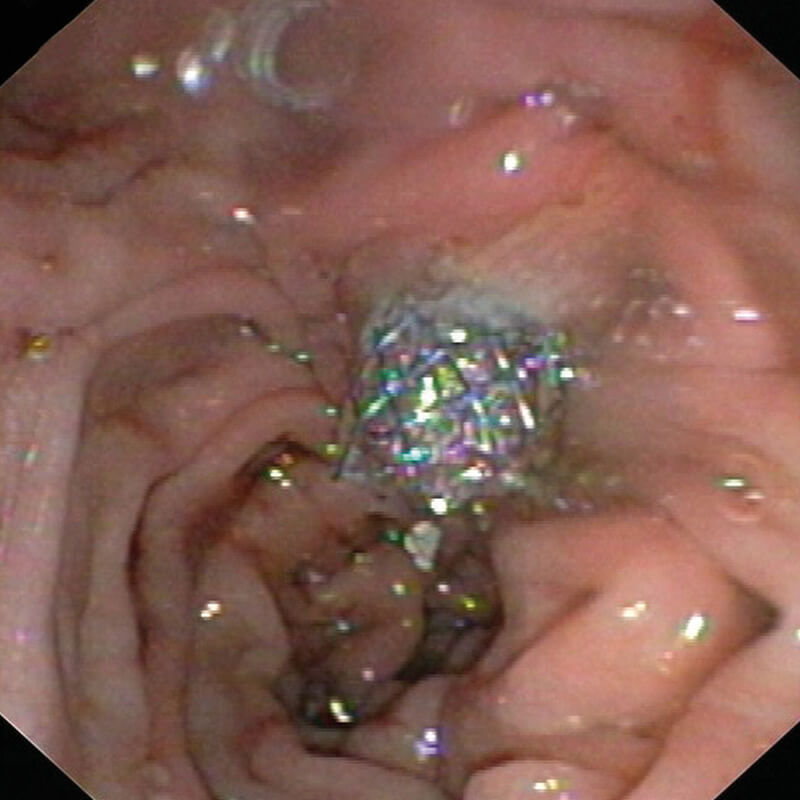
Endoscopic positional control
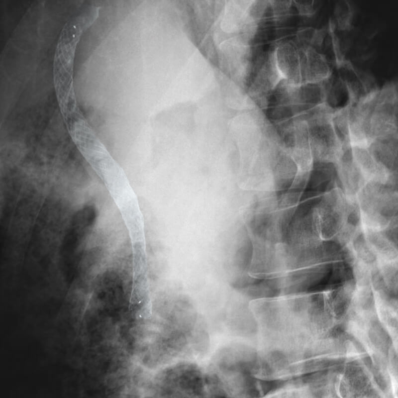
Released stent
CLASSIC-LINE. FIRST-CHOICE FOR ALL APPLICATIONS
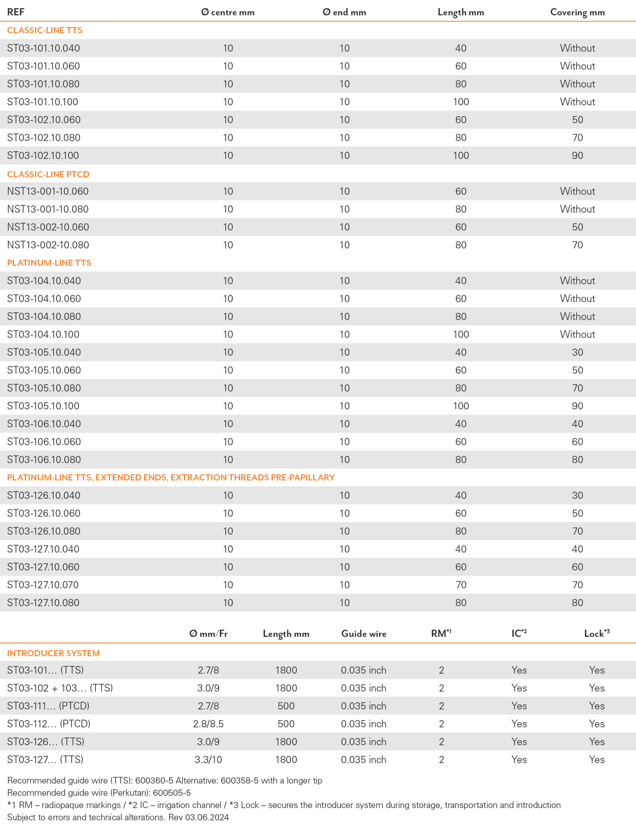
The Classic-Line is perfectly suited for all standard interventions. With two different approaches consisting of TTS and PTCD, it perfectly meets every requirement. These stents are made of highly flexible Nitinol wire mesh. Ten additional radiopaque markers account for the good visibility of our Classic-Line stents under fluoroscopy. Our product range comprises uncovered and partially covered stents.
PTCD FOR PERCUTANEOUS ACCESS
In case of therapeutic interventions into the biliary duct, it sometimes occurs that a transpapillary access cannot be accomplished. In such cases MICRO-TECH offers PTCD systems (percutaneous transhepatic cholangio drainage) as the ideal solution.
TTS STENTS FOR ROUTINE INTERVENTIONS
We offer uncovered as well as partially covered biliary duct stents in three different lengths. All stents can be placed through the working channel of the duodenoscope over the inserted guide wire (TTS – through-the-scope).
INTRODUCER SYSTEM FOR AN EXACT RELEASE
Additional radiopaque markers on the distal end of the introducer system and on the tip of the pusher catheter provide for additional orientation. In the endoscopic image, the tip of the pusher catheter can be unambiguously and easily distinguished from the proximal end of the stent. The permanent visual endoscopic control of the stent‘s proximal end considerably facilitates the exact release of the stent.
TEN PLATINUM RADIOPAQUE MARKERS
In order to achieve improved visibility under X-ray control each stent bares a total of ten radiopaque markers: four markers located at each end, and two in the middle of the stent. The two radiopaque markers in the middle guarantee for the controllability of the stent‘s optimal position during deployment.

